
MRI and CT
CT Scanning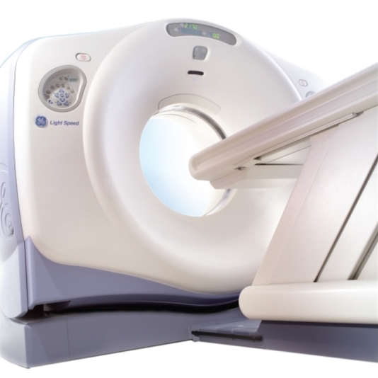
Computed Tomography (CT) imaging uses X–rays in conjunction with computing algorithms to image the body. In CT, an X–ray generating tube opposite an X–ray detector (or detectors) in a ring-shaped apparatus rotate around a patient producing a computer generated cross–sectional image (tomogram). Iodinated contrast agents are typically used with CT for enhanced delineation of anatomy and angiography. With computer manipulation, CT images can be reconstructed into two–dimensional and three–dimensional images. PetCure Oncology at VRIC uses a helical / spiral scanner to rapidly acquire images of dogs, cats and exotic animals. General anesthesia is used because most studies require the patient to remain motionless for a few minutes.
When complex radiosurgery planning is necessary for cancer treatment, CT imaging is used because it provides information about where the cancer is located and what vital structures are around the cancer. Precise localization of normal and abnormal tissues is important so that the targeted radiosurgery beam destroys abnormal cancer tissue, but spares normal tissues.
MRI ScanningMagnetic Resonance Imaging (MRI) uses strong magnetic fields to align spinning atomic nuclei (usually hydrogen protons) within body tissues, then uses a radio signal to disturb the axis of rotation of these nuclei and observes the radiofrequency signal generated as the nuclei return to their baseline states. The radio signals are collected by small antennae, called coils, placed near the area of interest. An advantage of MRI is its ability to produce images in axial, dorsal, sagital and multiple oblique planes with equal ease. MRI scans give the best soft tissue contrast of all the imaging modalities.
The center allows all veterinarians access to advanced cross-sectional imaging for their patients in an environment specifically designed for pets. Our equipment includes a 1.5 Tesla GE Signa Advantage MRI, which is the highest field strength MRI unit available for animals in the state. The MRI center uses specialized, MRI compatible patient monitoring and anesthesia delivery equipment. We use Sevoflurane gas anesthesia for the most rapid recovery and to minimize anesthetic complications for increased overall safety of your patients.
Equipment MaintenanceThe imaging equipment available at PetCure Oncology at VRIC was purchased from GE and is maintained in top working condition under service contract with Oxford Instruments OiS Technicians who also provide service in human imaging facilities in the area.
Image InterpretationA board–certified veterinary radiologist is available at the center to assist in determining the imaging modality of choice and provides the most accurate image interpretation possible. Written results of the imaging studies are provided to the referring veterinarian managing the patient's care within 24-48 hours of the imaging study. A digital copy of the imaging study is provided to the owner at the time they pick up their pet. Digital copies of the studies are transmitted to referring veterinarians and offsite hospitals when requested.